Low-level laser therapy
Low-level laser therapy, also known as photodynamic therapy
Low-level laser therapy involves the use of light radiation consisting of particles with wave properties called photons. Photons, by nature, are electromagnetic radiation with energy. When they interact with molecules and atoms, they can dislodge an electron from its orbit, resulting in the creation of a new photon and the release of energy.
Lasers emit monochromatic wavelengths, focusing relatively high-energy beams of light with a small scattering angle.
The effects of laser beams on living tissues occur in three ways:
- Thermal: They create a local heating effect.
- Mechanical: They generate vibrations similar to ultrasound in tissues.
- Biological: They act on the cellular level on existing enzymes, affecting their activity, etc.
Low-level laser therapy is widely used in medicine.
A low-level laser is a non-invasive light source that generates light of a specific wavelength without thermal, sound, or vibration effects, inducing biostimulation and photobiomodulation in tissues. As a result, pain is reduced, and cell stimulation occurs.
The effects of low-level laser therapy on living tissues include:
- Thermal and bioenergetic effects: Improved impulse conduction in nerve cells and expanded blood vessels.
- Biochemical effects: Enhanced synthesis of ATP, improved migration of fibroblasts, increased activity of macrophages, increased keratinocyte activity in the skin, promotion of RNA synthesis, and intensified enzyme formation.
- Bioelectrical effects: Improved operation of ion channels in cell membranes and changes in intracellular and extracellular ion gradients.
Clinical effects:
- Reduced muscle tension and pain; local blood circulation improves;
- Improved mobility of joints and connective tissues, increased tissue healing process;
Low-energy laser therapy is used to treat health problems such as carpal tunnel syndrome, fibromyalgia, osteoarthritis, rheumatoid arthritis, and non-healing wounds. This therapy is also used in dentistry to treat inflammation in the mucous membranes.
Physiological effects of laser therapy:
- Activation of immune cells, which promotes the fight against inflammation and improves tissue healing. The release of endorphins is promoted and ATF production in nerve cells is accelerated;
- Microcirculation improves and phagocytosis of leukocytes increases. As a result, the biopotential of cell membranes is stabilized. Stabilization of membranes reduces the feeling of pain;
- The release of inflammatory cytokines decreases (production of interleukin 1 – IL-1);
- Synthesis of prostaglandins, especially PG12, increases, which causes vasodilatation, reduces platelet aggregation and reduces inflammation.
- Ion transport in the cells improves – as sodium and calcium accumulate in them, ATF formation is disturbed;
- Restoration of energy metabolism of damaged cells;
- More energy is used for the synthesis of ATF;

Local Laser Therapy
Local laser therapy utilizes red, blue, green, and infrared light rays, each differing in wavelength and tissue penetration capability.
Blue laser light – with a wavelength of 405 nm, it can penetrate tissues to a depth of 1 – 2 mm. Blue light possesses antibacterial and anti-inflammatory properties, promoting the release of nitric oxide in tissues, thereby locally improving tissue microcirculation and stimulating mitochondria in cells, enhancing metabolism.
Green laser light – with a wavelength of 532 nm, it can penetrate tissues from 0.5 – 1 cm. It improves oxygen supply to tissues, beneficially affects blood vessel walls, normalizes the function of sodium-potassium pumps in erythrocyte membranes, and promotes mitochondrial activity.
Red laser light – with a wavelength of 658 nm, it can reach tissues from 2 – 3 cm deep. It enhances cell activity and improves microcirculation. By affecting specific types of leukocytes, it stimulates immune system activity, enhances fibroblast activity, promotes wound healing, and stimulates mitochondrial function.
Infrared radiation – with a wavelength of 810 nm, its penetration capability in tissues ranges from 5 – 7 cm. By stimulating mitochondria, it promotes the release of ATP in cells, locally improving metabolic processes.
Intravenous or endovascular photodynamic therapy
Intravenous laser therapy has been used as a medical method since 1981. Initially, it was used to improve blood rheological properties in patients after myocardial infarction.
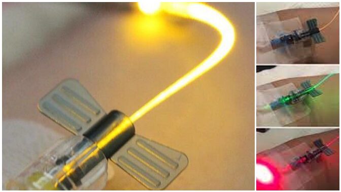
It is performed with low-energy laser beams (1 – 5 mW) for a duration of 20 – 60 minutes. The average course of therapy is 10 sessions, performed daily or three times a week. During these procedures, an intravenous cannula is inserted into the elbow or wrist vein, followed by the attachment of a catheter and a light source.
Photosensitizing Agents
Photosensitizing agents are used in intravenous laser therapy to facilitate the transfer of light energy. They have properties to absorb light waves of a specific wavelength and then transfer them to adjacent molecules. These agents have selectivity for specific tissues, where they accumulate and transfer light energy to cells. By activating mitochondrial membranes within cells, they promote intracellular programmed cell death or apoptosis. Therefore, these agents are widely used in integrative oncology.
Photosensitizing agents are typically water-soluble for easier transportation throughout the body. They themselves are non-toxic and do not produce toxic metabolites. They are most commonly derived from plant extracts.
Photosensitizing agents with a structure similar to porphyrins include derivatives of hematoporphyrin, benzoporphyrin, texaphyrin, 5-aminolevulinic acid, and chlorine E-6.
- Aminolevulinic acid (ALA) absorbs red laser beams with a wavelength of 630 nm. It is used in dermatology for the treatment of various skin diseases, as well as skin cancer, bladder cancer, superficial tumors of the head, neck, and oral cavity.
- Chlorin E-6 is widely used in tumor photodynamic therapy. This substance is obtained from green algae found in nature. It absorbs red light beams with a wavelength of 660 nm very effectively. During therapy, it binds to tumor cells with great precision and promotes their destruction. It is completely eliminated from the body within 48 hours. Just a few hours after photodynamic therapy, chlorine E6 begins apoptosis of tumor tissues and induces cell necrosis. Chlorine E6 is also used in diagnostic purposes with blue light at a wavelength of 405 nm to specify the localization of tumor tissues. Other similar chlorine-based substances that are used include chlorins, purpurins, and bacteriochlorins.
- Curcumin is also commonly used in photodynamic therapy. It is derived from the Curcuma longa plant, widely used as a spice. Curcumin absorbs blue light with a wavelength of 440 nm even at low concentrations. It is very effective in the treatment of psoriasis, tumors, various chronic infections, and other diseases. It possesses apoptosis-inducing, anti-inflammatory, immune-stimulating, and anticancer properties. Currently, curcumin can be obtained as a purified infusion solution, which binds very well to plasma albumins in the blood.
- Hypericin is a photosensitive substance of plant origin obtained from St. John’s wort. It absorbs yellow light with a wavelength of 589 nm very well. St. John’s wort extract consists of hypericin, pseudohypericin, and hyperforin. Currently, there is a 10 mg vial of purified hypericin available in pharmacies for intravenous administration, which binds well to plasma albumins. Hypericin is used in the treatment of tumors, depression, as well as chronic viral and bacterial infections.
- Epigallocatechin gallate (EGCG) is the latest photosensitizing agent, which is structurally a green tea polyphenol. It absorbs red light at a wavelength of 662 nm. Photodynamic therapy is used in the treatment of various types of tumors.
- Riboflavin, also known as vitamin B2, is present in all aerobic organisms, as well as in several foods such as milk, yeast, beer, eggs, and leafy vegetables. It is considered a very safe photosensitizing agent. It absorbs visible light at a wavelength of 446 nm. This substance is also formed in small amounts in the human body itself and is neither toxic nor mutagenic. From riboflavin, substances are formed in the body which later act as coenzymes and participate in cellular metabolic processes.
Biological Effects of Intravenous Laser Therapy:
- Normalization of cell membrane biopotential;
- Activation of specific and nonspecific immune responses;
- Increase in IgG, IgM, and IgA immunoglobulin levels in the blood;
- Lymphocyte proliferation;
- Increase in interleukins, interferons, and TNF alpha levels in the blood;
- Enhanced macrophage phagocytic activity;
- Reduction of inflammatory indicators in the body;
- Improved antioxidant and detoxifying effects;
- Promotion of erythrocyte regeneration and microcirculation;
- Reduction in platelet aggregation;
- Activation of fibrinolysis;
- Increased production of nitric oxide, promoting vasodilation and preventing endothelial dysfunction;
- Stimulation of ATP production in mitochondria, improving metabolism.

The use of intravenous laser therapy or photodynamic therapy:
- In sports medicine;
- Treatment of autoimmune, neurodegenerative, and chronic inflammatory diseases;
- Treatment of coronary heart disease;
- Treatment of fibromyalgia;
- Treatment of tumors;
- Treatment of chronic gastrointestinal diseases;
- Treatment of chronic viral and bacterial infections;
- Treatment of depression, Alzheimer’s disease, and dementia;
- Treatment of skin diseases, etc.
